
DXA Bone Densitometer DEXA Pro-1
Application
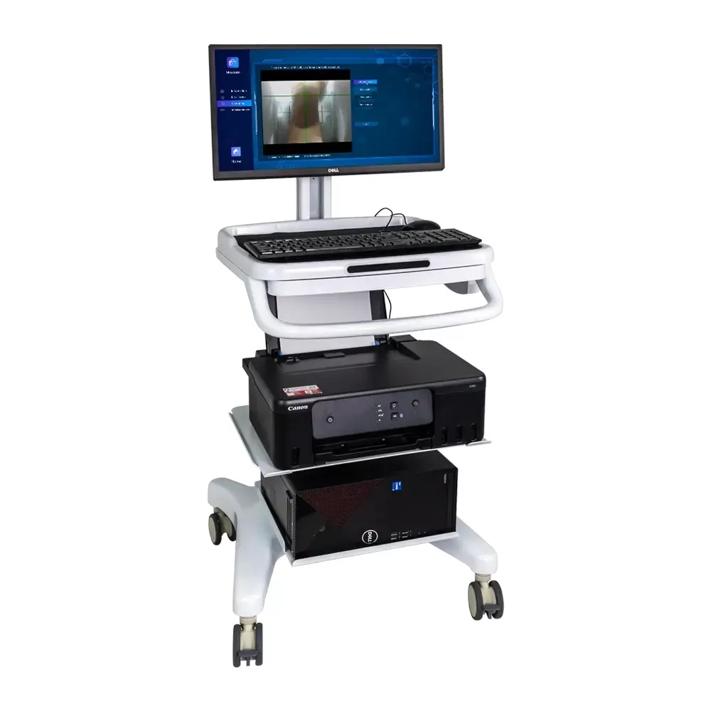
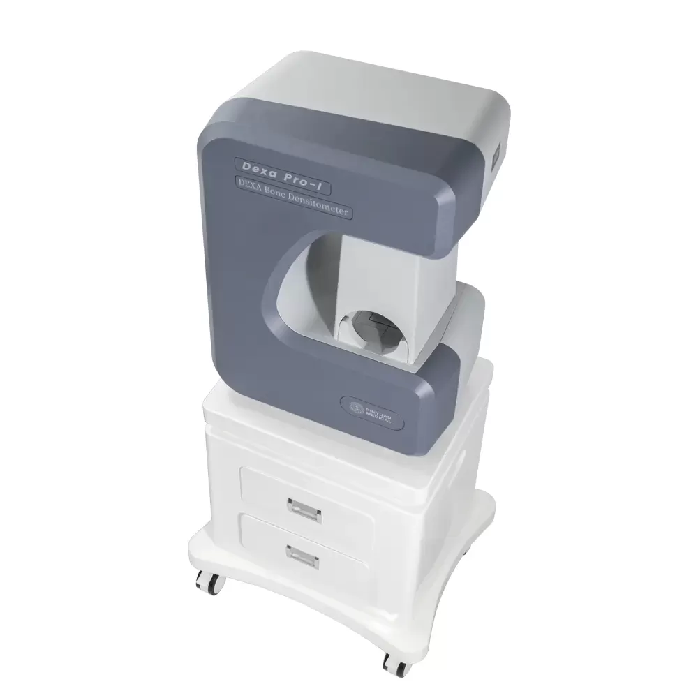
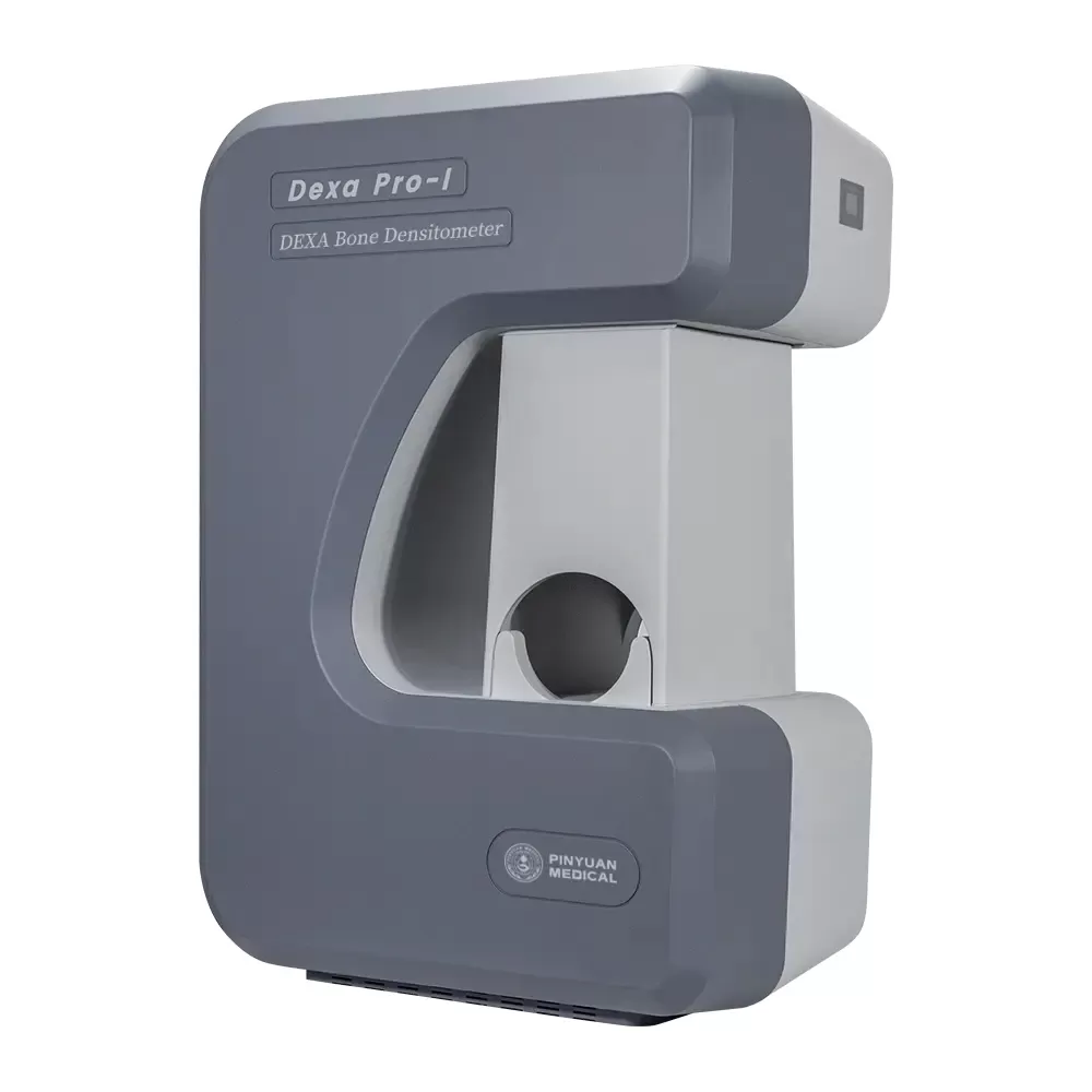
Dual-Energy X-ray Absorptiometry (DXA or DEXA) useing a very small dose of ionizing radiation to produce pictures of the inside of the forearm to measure bone density . It is evaluating for Osteoporosis and Osteopenia and provides a versatile solution for evaluating the risk of osteoporotic fracture.
It is an enhanced form of X-ray technology that is used to measure bone loss. DXA is today's established standard for measuring bone mineral density (BMD).
Technical Parameter
1.Using the Dual Energy X-ray Absorptimetry.
2.Using the Most Advanced Cone - Beam and Surface Imaging Technology.
3.With High Measurement Speed and Short Measurement Time.
4.With Dual Imaging Technology to Get More Accurate Measurement.
5.Using Laser Beam Positioning Technique, Making the Measuring Position More Accurate.
6.Dectcing Image Digitization, to Get Accurate Measurement Results.
7.Adopting the Surface Imaging Technology, Measuring Faster and Better.
8.Using the Unique Algorithms to Get More Accurate Measurement Results.
9.Adopting the Full Closed Lead Protective Window to Measure, only Need to Put the Patient's Arm into the Window. The Equipment is Indirect Contact with the Scanning Parts of the Patient. Easy to Operate for the Doctor. It is Safety for the Patient and Doctor.
10.Adopting Integrated Structure Design
11.Unique Shape, Beautiful Appearance and Easy to Use.
Performance Parameter
1.Measurement Parts: the Front of Forearm.
2. X ray tube voltage:High Energy 85Kv, Low Energy 55Kv.
3.The high and low energy corresponds to the current, 0.2mA at high energy and 0.4mA at low energy
4.X-Ray Detector:Imported High Sensitivity Digital Camera.
5.X-Ray Source:Stationary Anode X-ray Tube (with High Frequency and Small Focus)
6.Imaging Way:Cone - Beam and Surface Imaging Technology.
7.Imaging Time:≤ 5 Seconds.
8.Accuracy(error)≤ 1.0%
9.Repeatability Coefficient of Variation CV≤0.5%
10.Can be connected to hospital HIS system, PACS system
11.Measuring Parameter: T- Score, Z-Score, BMD,BMC, Area,Adult percent[%], Age percent[%], BQI (The Bone Quality Index) ,BMI,RRF: Relative Fracture Risk
12. It with multi race clinical database, including: European, American, Asian, Chinese, WHO international compatibility. It measuring the people between the age of 0 and 130.
13.Original Dell Business Computer: ,Intel i5,Quad Core Processor ,8G,1T,22'inch HD Monitor
14.Operation System: Win7 32-bit / 64 bit ,Win10 64 bit compatible
15.Working Voltage: 220V±10%, 50Hz.
What Is a Bone Mineral Density Test?
A bone mineral density test, sometimes just called a bone density test, detects whether you have osteoporosis, a word that comes from Greek and literally means "porous bone."
When you have this condition, your bones get weak and thin. They become more likely to break. It's a silent condition, which means you don't feel any symptoms. Without a bone density test, you may not realize you have osteoporosis until you break a bone.
How the Test Works
The bone density test is painless and quick. It estimates how dense or thick your bones are by using X-rays.
The DXA Bone Densitometry 500A measure how much calcium and minerals are in a part of your bone. The more minerals you have, the better. That means your bones are stronger, denser, and less likely to break. The lower your mineral content, the greater your chance of breaking a bone in a fall.
FAQs for DXA 500A Forearm Bone Densitometer
1. Q: The device uses X-rays. How is safety ensured for both patients and my staff in a clinic without a lead-lined room?
A: Safety is engineered into the core design. The DXA 500A features a fully enclosed lead protective window. The patient only places their forearm inside this shielded window during the ≤5-second scan. Combined with remote console operation, this design contains radiation completely, eliminating any need for a dedicated, expensive lead-lined room and ensuring a safe environment for operators.
2. Q: Why measure only the forearm? Is this sufficient for a reliable osteoporosis diagnosis?
A: Forearm DXA is a well-validated and powerful screening tool. Measurement of the radius and ulna is highly sensitive to age-related bone loss and strongly correlates with overall fracture risk. With excellent precision (CV≤0.5%), it is ideal for high-volume population screening and for monitoring changes in bone density over time. For cases identified as high-risk, a follow-up central (spine/hip) DXA can be recommended, making this a highly efficient and cost-effective diagnostic pathway.
3. Q: We serve a multi-ethnic population. How does the device ensure accurate T-scores for all our patients?
A: This is a critical feature. The DXA 500A includes multiple, validated reference databases (European, American, Asian, Chinese). The system can select the appropriate ethnic-specific database for each patient. This ensures the T-scores and Z-scores are calculated using the correct normative data, providing accurate and clinically meaningful results for your diverse patient base and preventing misdiagnosis.
4. Q: What is the practical benefit of "Cone-Beam and Surface Imaging Technology"?
A: This advanced dual-imaging approach delivers both speed and superior accuracy. Cone-beam imaging enables rapid scanning (≤5 seconds), improving patient comfort and clinic throughput. Simultaneously, surface imaging provides sharper bone edge detection, leading to more precise bone area definition and, consequently, more accurate and reproducible BMD calculations.
5. Q: Can this device integrate with our hospital's existing IT infrastructure (HIS/PACS)?
A: Yes, seamlessly. The DXA 500A is designed for compatibility with standard Hospital Information Systems (HIS) and Picture Archiving Systems (PACS). This allows for efficient patient data management, automatic transfer of reports and images to electronic medical records, and streamlined workflow within your clinical network.
6. Q: The report includes BQI and RRF. What are these, and how do they add value beyond the standard T-score?
A: These parameters offer a more comprehensive bone health assessment. BQI (Bone Quality Index) provides insights into bone microstructure and strength beyond mere mineral density. RRF (Relative Fracture Risk) offers a quantified risk assessment. Together with the T-score, they give clinicians a multi-dimensional view to support more personalized patient management and treatment decisions.
7. Q: How easy is it for our clinical staff to operate? Does it require a specialized radiographer?
A: Operation is simplified for efficiency. The laser beam positioning guide and intuitive software interface make patient setup quick and accurate. Comprehensive training is provided, enabling nurses, technicians, or physicians to become proficient operators rapidly, reducing reliance on specialized radiology staff.
8. Q: You specify an original Dell business computer. Why is this important?
A: Including a high-quality, brand-name workstation (Dell, Intel i5, 8GB RAM) guarantees system stability, data security, and reliable long-term performance. It ensures the sophisticated imaging software runs smoothly, protects patient data, and provides access to professional technical support, safeguarding your investment and daily operations.
9. Q: What is the total cost of ownership (TCO) advantage compared to a traditional whole-body DXA system?
A: The TCO advantage is substantial. The DXA 500A eliminates the high cost and regulatory hassle of building a lead-lined room. Its purchase price, footprint, and operational power consumption are significantly lower. This makes gold-standard DXA technology accessible and financially viable for community hospitals, outpatient clinics, and specialized practices.
10. Q: Beyond basic osteoporosis screening, what are other applications for this device?
A: Its applications are versatile:
Treatment Monitoring: Precisely track the efficacy of osteoporosis medications over time.
Pediatric & Adolescent Health: Assess bone development and peak bone mass acquisition using age-matched Z-scores.
Clinical Research: An excellent tool for epidemiological studies due to its speed, precision, and portability.
Specialist Management: Ideal for endocrinology, rheumatology, and geriatrics departments to manage patients with secondary osteoporosis risks.
Popular Products
Ultrasound Bone Densitometer BMD-A1 Basic Osteoporosis Detector
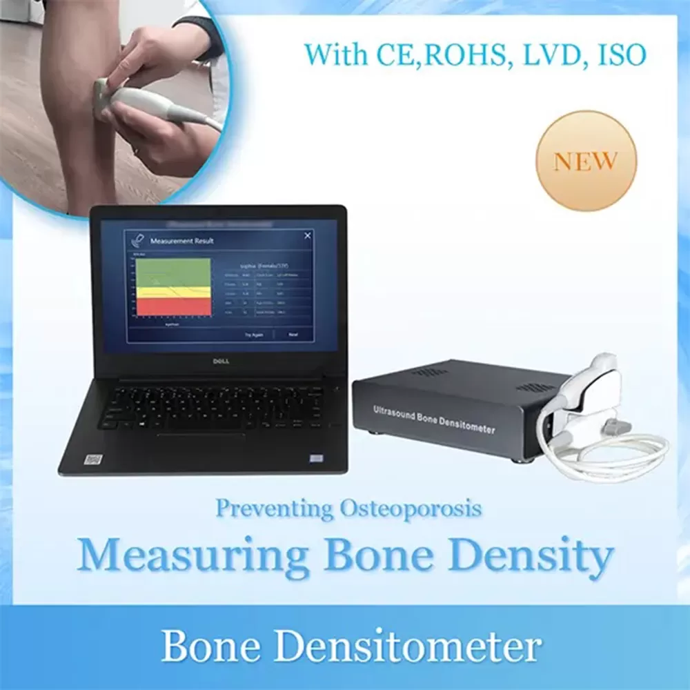
Portable Ultrasound Bone Densitometry BMD-A1 Osteoporosis Detector
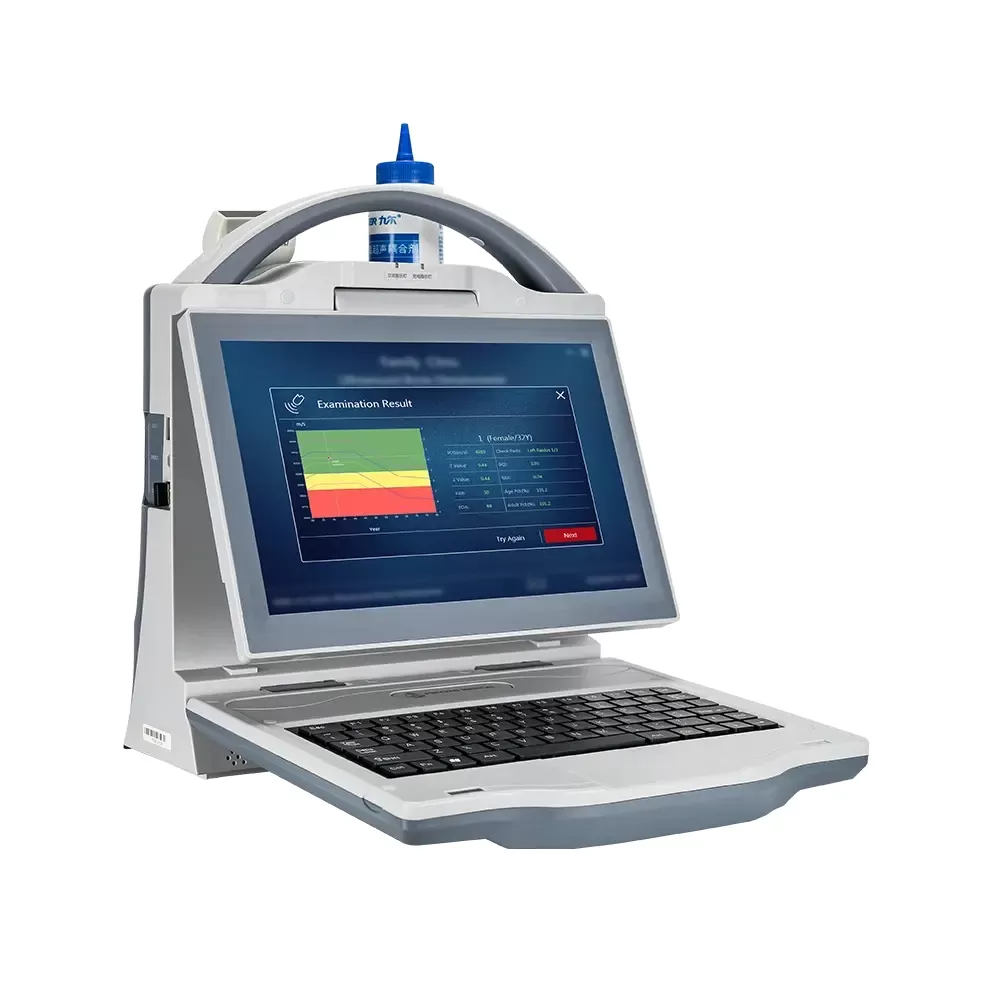
Portable Ultrasound Bone Densitometer BMD-A3
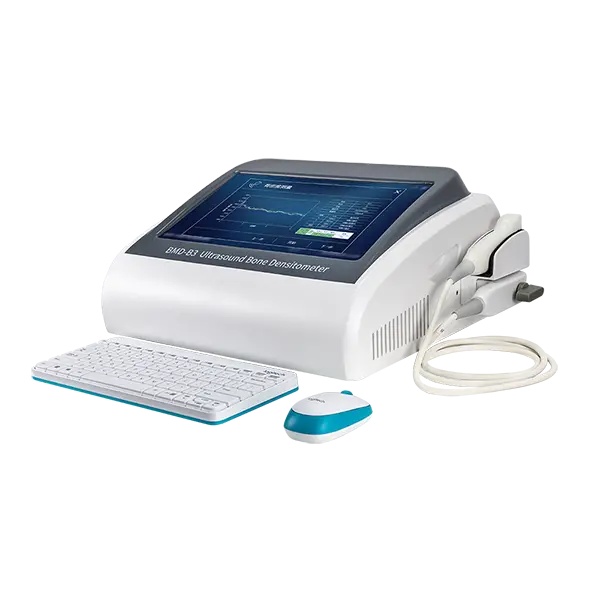
Automatic High Effective Portable Ultrasound Bone Densitometer BMD-B3
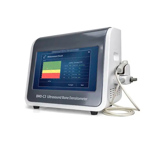
Portable Ultrasound Bone Densitometer BMD-C3
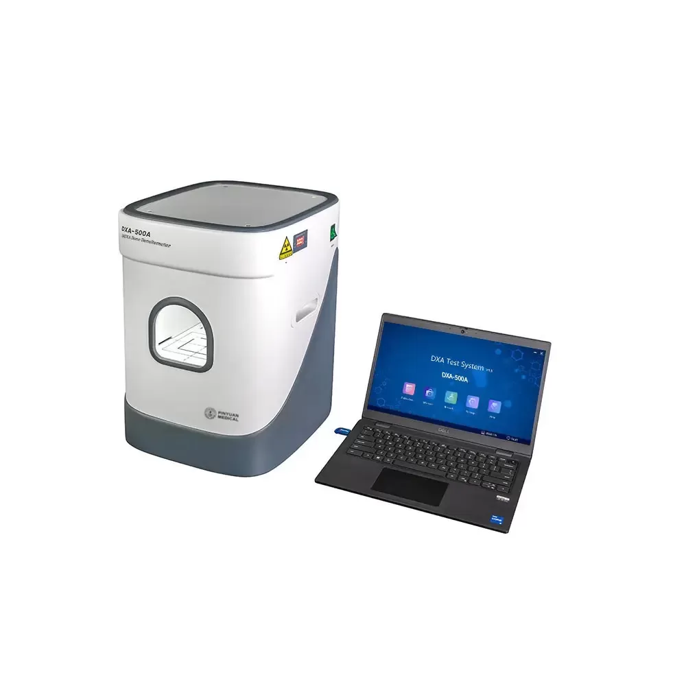
DXA Bone Densitometer 500A Dual-Energy X-ray Absorptiometry
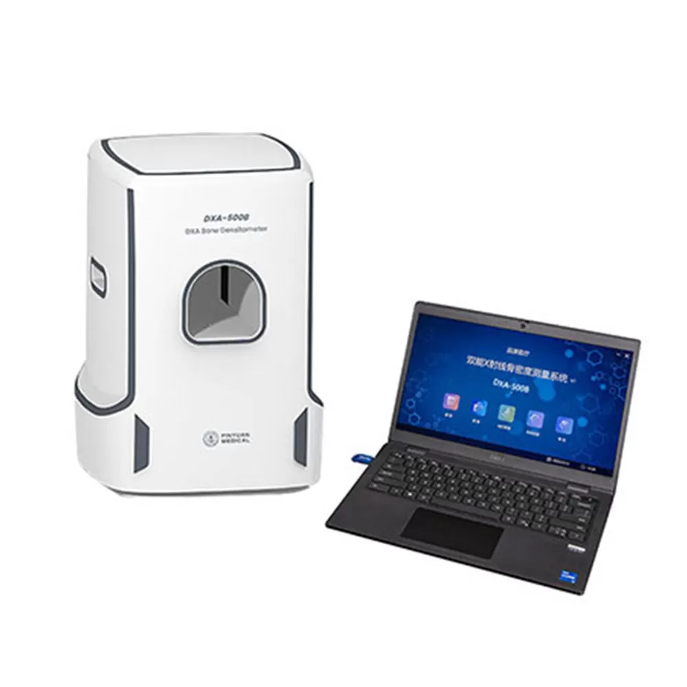
Dual-Energy X-ray Bone Densitometer DXA-500B
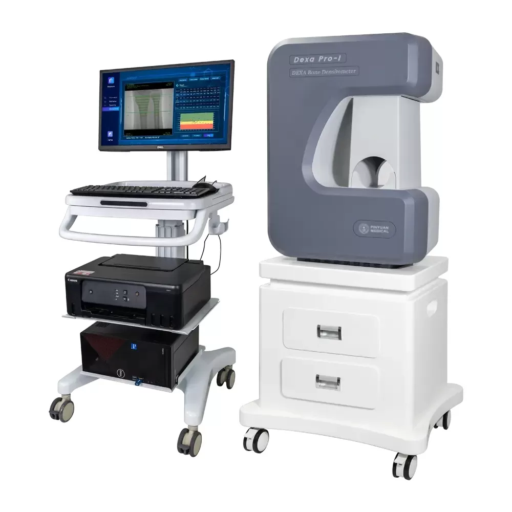
DXA Bone Densitometer DEXA Pro-1
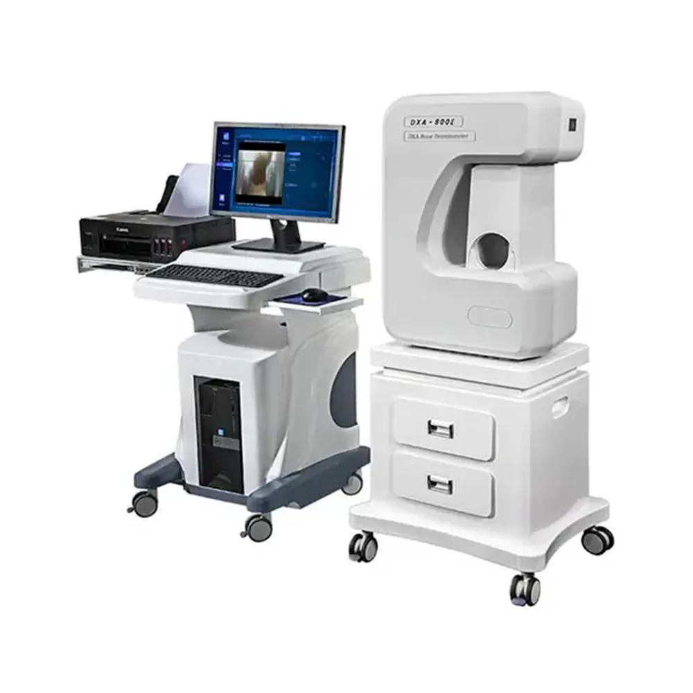
DXA Bone Densitometer 800E
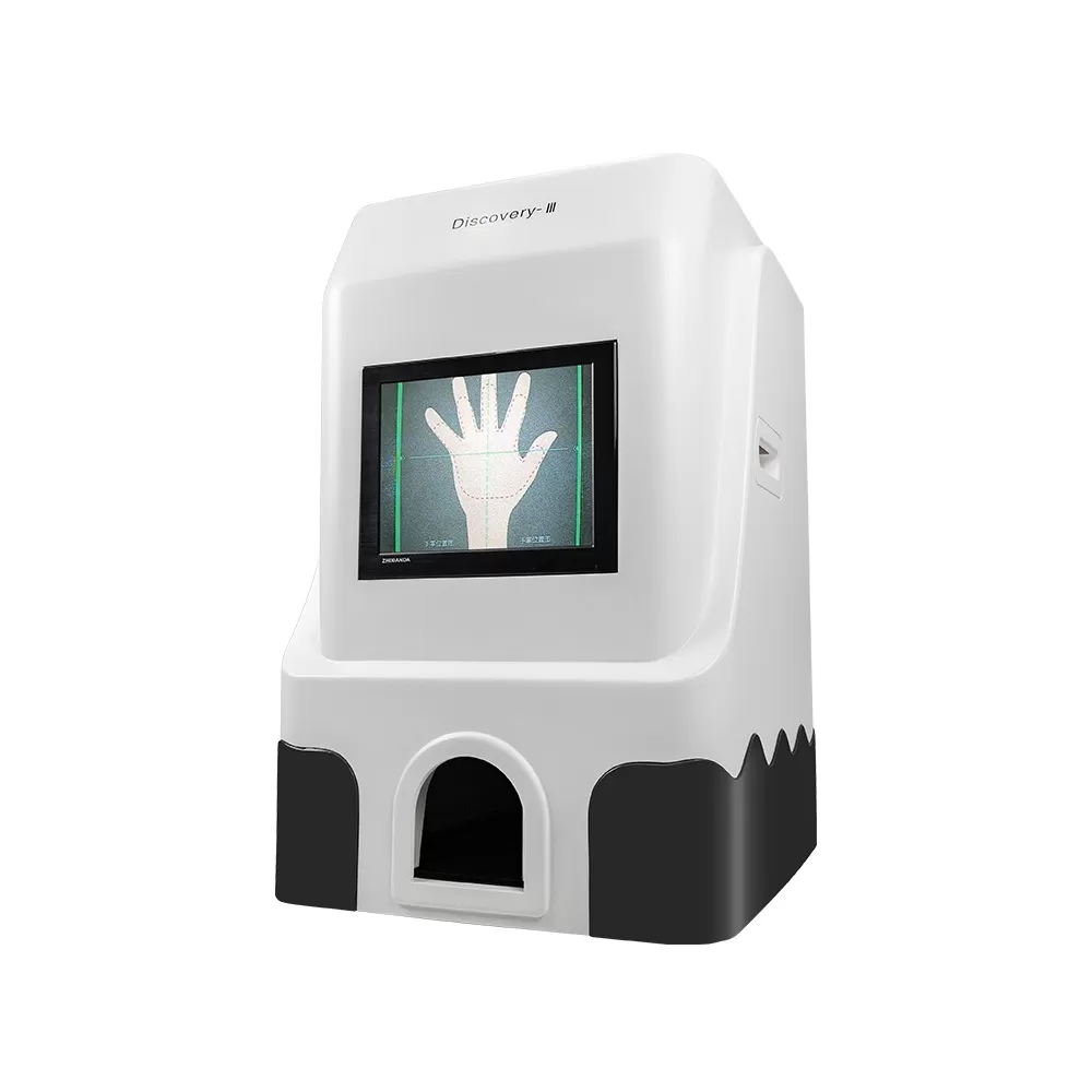
X-ray Bone Density and Skeletal Age Instrument Discover-lll