
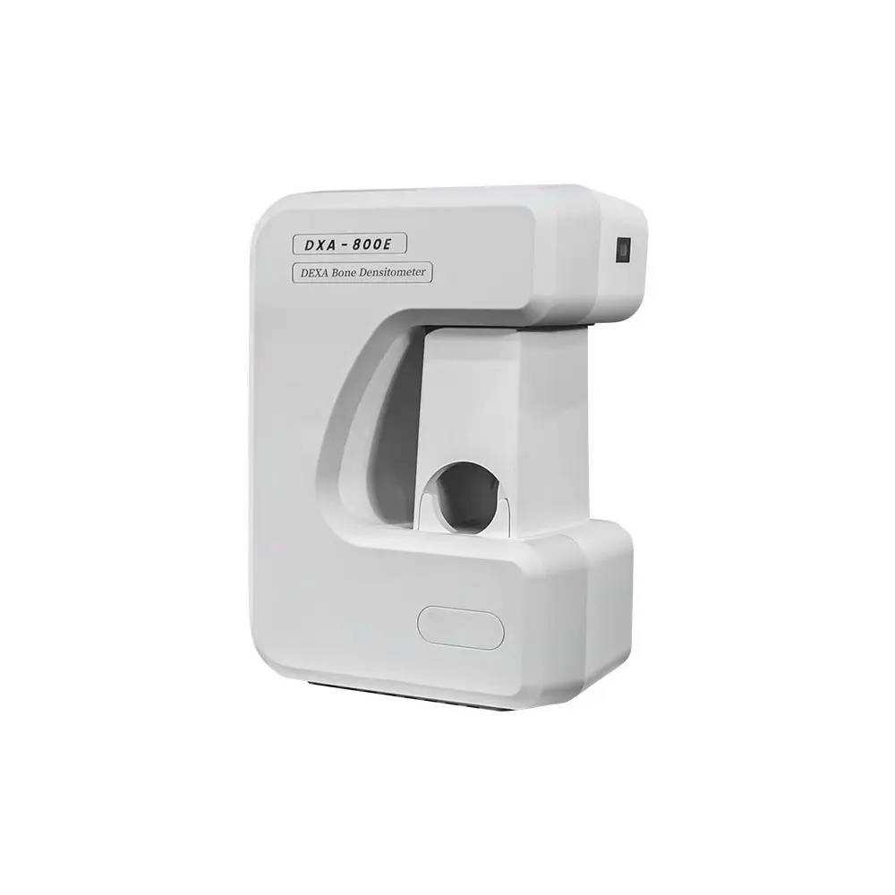
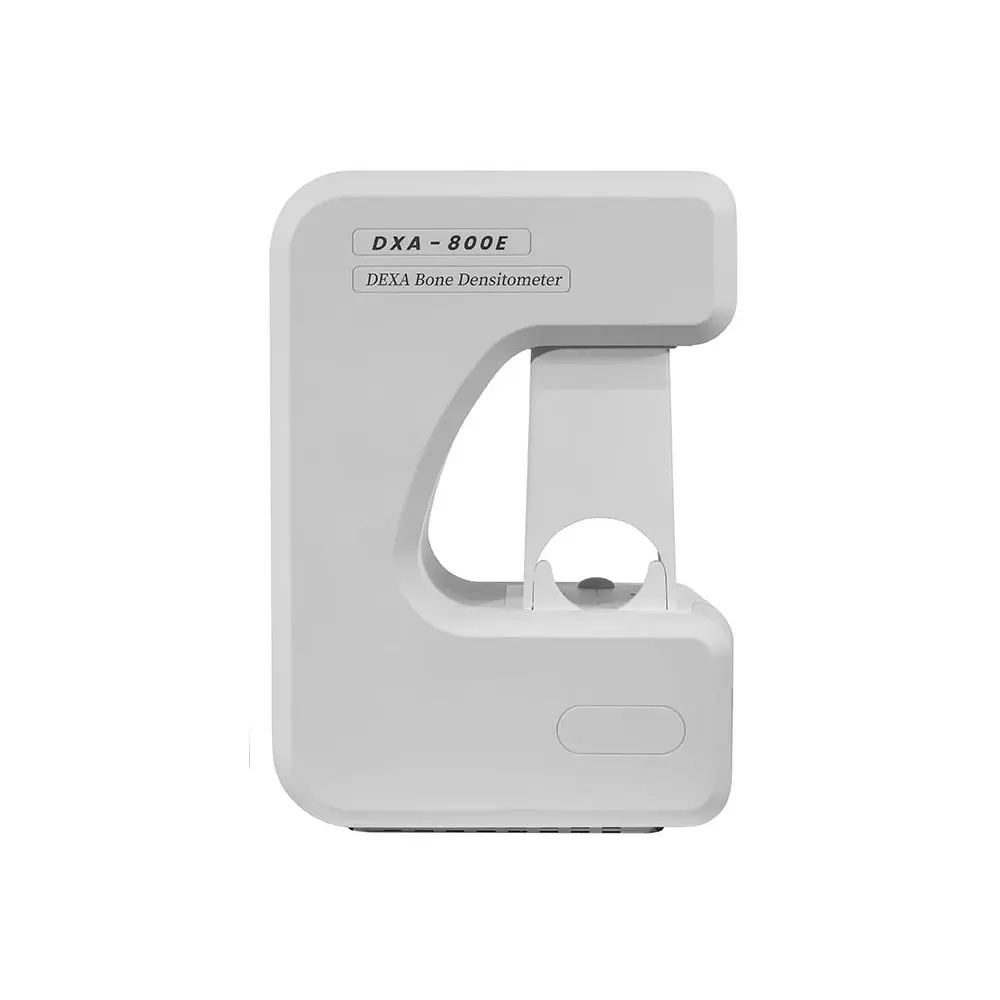
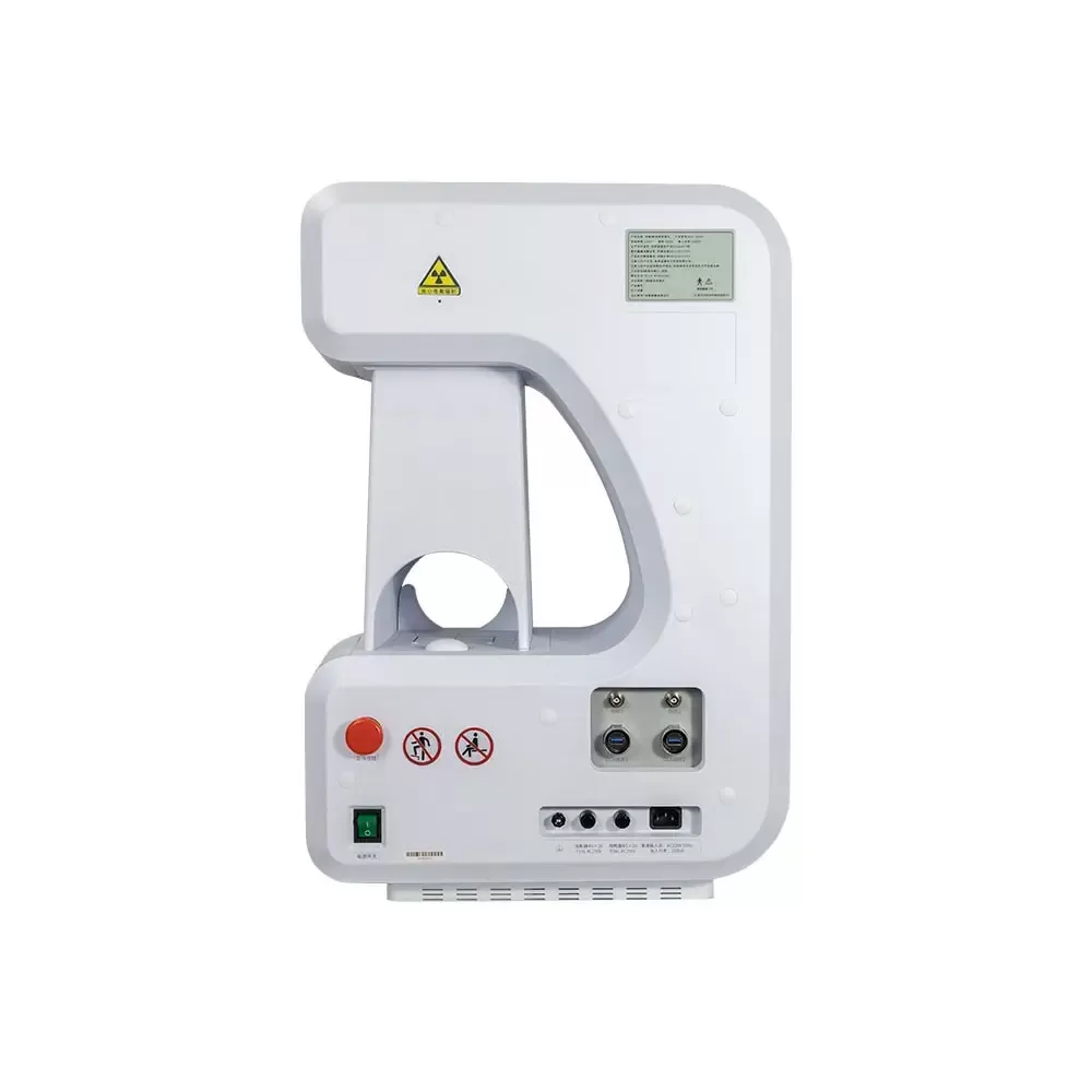
DXA Bone Densitometer 800E
Application
Dual-Energy X-ray Absorptiometry (DXA or DEXA) using a very small dose of ionizing radiation to produce pictures of the inside of the forearm to measure bone density . It is evaluating for Osteoporosis and Osteopenia and provides a versatile solution for evaluating the risk of osteoporotic fracture.
It is an enhanced form of X-ray technology that is used to measure bone loss. DXA is today's established standard for measuring bone mineral density (BMD).
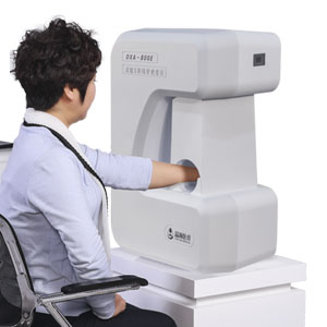
Features
Using digital Laser Beam Positioning Technique.
Special Analysis System Based on Different Countries People.
Using the Most Advanced Cone - Beam and Surface Imaging Technology.
Measurement Parts: the Front of Forearm.
With High Measurement Speed and Short Measurement Time.
Adopting the Full Closed Lead Protective Window to Measure.
Technical Specifications
Large Scale Integrated Circuit.
Multi -Layer Circuit Board Design.
Light Source Technology With High Frequency and Small Focus.
Imported High Sensitivity Digital Camera.
Using the Cone - Beam and Surface Imaging Technology.
Using Laser Beam Positioning Technique.
Using the Unique Algorithms.
ABS Mould Manufactured, Beautiful, Strong and Practical.
Special Analysis System Based on Different Countries People.
Technical Parameter
1.Using the Dual Energy X-ray Absorptimetry.
2.Using the Most Advanced Cone - Beam and Surface Imaging Technology.
3.With High Measurement Speed and Short Measurement Time.
4.With Dual Imaging Technology to Get More Accurate Measurement.
5.Using Laser Beam Positioning Technique, Making the Measuring Position More Accurate.
6.Dectcing Image Digitization, to Get Accurate Measurement Results.
7.Adopting the Surface Imaging Technology, Measuring Faster and Better.
8.Using the Unique Algorithms to Get More Accurate Measurement Results.
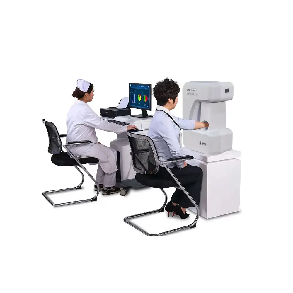
9.Adopting the Full Closed Lead Protective Window to Measure, only Need to Put the Patient’s Arm into the Window. The Equipment is Indirect Contact with the Scanning Parts of the Patient. Easy to Operate for the Doctor. It is Safety for the Patient and Doctor.
10.Adopting Integrated Structure Design.
11.Unique Shape, Beautiful Appearance and Easy to Use.
Performance Parameter
1.Measurement Parts: the Front of Forearm.
2. X ray tube voltage: High Energy 70 Kv, Low Energy 45Kv.
3.The high and low energy corresponds to the current, 0.25 mA at high energy and 0.45mA at low energy.
4.X-Ray Detector: Imported High Sensitivity Digital Camera.
5.X-Ray Source: Stationary Anode X-ray Tube (with High Frequency and Small Focus).
6.Imaging Way: Cone - Beam and Surface Imaging Technology.
7.Imaging Time: ≤ 4 Seconds.
8.Accuracy(error)≤ 0.4%.
9.Repeatability Coefficient of Variation CV≤0.25%.
10.Measuring Area :≧150mm*110mm.
11.Can be connected to hospital HIS system, PACS system.
12.Provide Worklist Port with independent upload and download function.
13.Measuring Parameter: T- Score, Z-Score, BMD, BMC, Area, Adult percent[%], Age percent[%], BQI (The Bone Quality Index) ,BMI、RRF: Relative Fracture Risk.
14. It with multi race clinical database, including: European, American, Asian, Chinese, WHO international compatibility. It measuring the people between the age of 0 and 130.
15.Measuring children over three years older.
16.Original Dell Business Computer: Intel i5, Quad Core Processor \8G \1T \22’inch HD Monitor.
17.Operation System: Win7 32-bit / 64 bit ,Win10 64 bit compatible.
18.Working Voltage: 220V±10%, 50Hz.
Who Should Get Bone Density Tested
Anyone can get osteoporosis. It’s more common among older women, but men can have it, too. Your chances increase as you age.
● You should discuss with your doctor whether you need the DXA Bone Test. They may recommend it if you meet any of the following:You’re a woman 65 or older.
● You’re a postmenopausal woman 50 or older.
● You’re a woman at the age of menopause and have a high chance for breaking bones.
● You’re a woman who has already been through menopause, younger than 65, and have other things that give you a higher chance of osteoporosis.
● You’re a man 50 or older with other risk factors.
● You break a bone after 50.
● You’ve lost more than 1.5 inches of your adult height.
● Your posture has gotten more hunched.
● You’re having back pain without any cause.
● Your periods have stopped or are irregular although you’re neither pregnant nor menopausal.
● You’ve gotten an organ transplant.
● You’ve had a drop in hormone levels.
Some types of prescription drugs can cause bone loss. These would include glucocorticoids, a class of drugs used to reduce inflammation. Tell your doctor if you’ve been on cortisone (Cortone Acetate), dexamethasone (Baycadron, Maxidex, Ozurdex), or prednisone (Deltasone).
Our DXA Bone Densitometry is for the Peripheral test. This measures bone density at your the front of forearm. It is usually cheaper.
Peripheral tests are also a way to screen people, so those who show a greater chance for osteoporosis can get more testing. They are also used for larger people who cannot get the central DXA because of weight limits.
The results on the bone density report
T score: This compares your bone density with a healthy, young adult of your gender. The score indicates if your bone density is normal, below normal, or at the levels that indicate osteoporosis.
Here’s what the T score means:
● -1 and above: Your bone density is normal.
● -1 to -2.5: Your bone density is low, and it may lead to osteoporosis.
● -2.5 and above: You have osteoporosis.
Z score: This allows you to compare how much bone mass you have compared with other people of your age, gender, and size.
FAQs for DXA Bone Densitometer
1. Q: The updated specs show improved accuracy (error ≤0.4%) and repeatability (CV≤0.25%). What does this mean for patient care?
A: These enhanced metrics signify top-tier precision in clinical measurement. The lower error margin ensures each single reading is exceptionally accurate, while the superior repeatability means you can trust the device to produce highly consistent results over time. This is crucial for confidently detecting subtle bone density changes, making it an extremely reliable tool for diagnosis, initial screening, and sensitive monitoring of treatment efficacy.
2. Q: The "Special Analysis System Based on Different Countries People" is mentioned again. How does it function in practice with the global databases?
A: This system is the software intelligence that leverages the multi-ethnic databases. When you input a patient's demographic data, it automatically selects and applies the correct reference curve (European, American, Asian, Chinese). This process ensures the calculated T-score and Z-score are ethnically calibrated, providing a fair and accurate assessment that accounts for physiological differences in bone density across populations, which is critical for avoiding misdiagnosis.
3. Q: The "Full Closed Lead Protective Window" is a key feature. Does this design have any operational drawbacks?
A: On the contrary, its operational benefits are significant. The design eliminates the need for direct contact between the device and the patient's skin (only the arm enters the window), streamlining infection control. It dramatically simplifies the procedure for the operator—just position and scan—while guaranteeing safety. There is no compromise in workflow; it only enhances it by integrating safety directly into the user experience.
4. Q: With an imaging time of ≤4 seconds, how does the "Cone-Beam and Surface Imaging Technology" achieve this speed without sacrificing image quality?
A: This dual-technology approach is key. Cone-beam imaging captures a volumetric data set in a single, rapid rotation. Simultaneously, surface imaging adds detailed topological information. The combination allows the system to acquire all necessary data in one swift pass, reducing motion artifact and patient discomfort, while the advanced processing algorithms ensure the extracted bone map and subsequent BMD calculation remain of the highest quality.
5. Q: The device can connect to hospital HIS/PACS and has a "Worklist Port." How does this improve clinic management?
A: This transforms the device from a standalone tool into an integrated node in your hospital's network. The Worklist Port allows it to automatically receive and manage patient queues from the HIS, eliminating manual data entry. After scanning, reports and images can be seamlessly uploaded to the PACS and EMR. This end-to-end digital integration minimizes administrative errors, saves time, and creates a smooth, efficient digital workflow.
6. Q: Who is the ideal candidate for this peripheral (forearm) DXA test versus a central (spine/hip) DXA?
A: This peripheral DXA is the ideal first-line screening tool for the broad risk groups listed (e.g., postmenopausal women, men over 50 with risk factors, individuals on steroids). It's perfect for high-volume screening programs, routine monitoring, and for patients who are ineligible for central DXA due to weight limits, mobility issues, or prior surgery. It efficiently identifies those who need the more comprehensive central DXA for definitive diagnosis.
7. Q: You mention it measures children over three years old. What is the clinical application in pediatrics?
A: In pediatrics, the focus shifts from diagnosing osteoporosis to assessing bone health development and detecting low bone density for age. Using the Z-score (comparison to age, gender, and size-matched peers), it helps diagnose conditions like juvenile osteoporosis, monitor bone health in chronic illnesses (e.g., rheumatoid arthritis, cystic fibrosis), and assess the impact of long-term medication use (e.g., glucocorticoids). It's a safe, low-dose tool for pediatric endocrinologists and rheumatologists.
8. Q: What is the practical advantage of the "Laser Beam Positioning Technique"?
A: It provides visual, real-time guidance for precise and reproducible patient positioning. The laser line shows exactly where to place the forearm, ensuring consistency across different operators and follow-up visits. This minimizes operator-dependent variability, which is a common source of error in bone densitometry, thereby guaranteeing that measured changes in BMD reflect true biological change, not positioning differences.
9. Q: The included computer is a branded Dell with an Intel i5 processor. Why is this level of specification important for a medical device?
A: This specification guarantees system stability, data processing speed, and long-term reliability. The bone density algorithms and image processing are computationally intensive. A professional-grade workstation ensures fast report generation, smooth software operation, and secure patient data storage, preventing crashes or slowdowns that could disrupt clinical workflow. It protects your investment by ensuring the hardware matches the sophistication of the medical software.
10. Q: For a private clinic or a community health center considering this device, what is the main value proposition?
A: The DXA 500A delivers "gold-standard" DXA technology in an accessible, turnkey package. It removes the major barriers of costly room shielding and high upfront investment associated with full-body systems. You receive a device with excellent accuracy, fast throughput, built-in safety, and professional reporting that enables you to offer a premium, in-house diagnostic service for osteoporosis management, creating a new revenue stream and enhancing patient care.
Popular Products
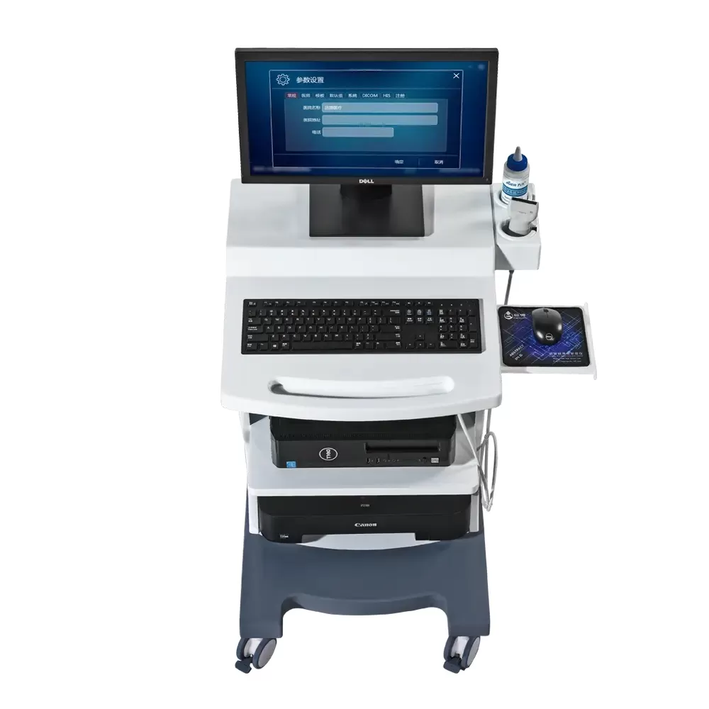
Ultrasound Bone Densitometer BMD-A1 Basic Osteoporosis Detector
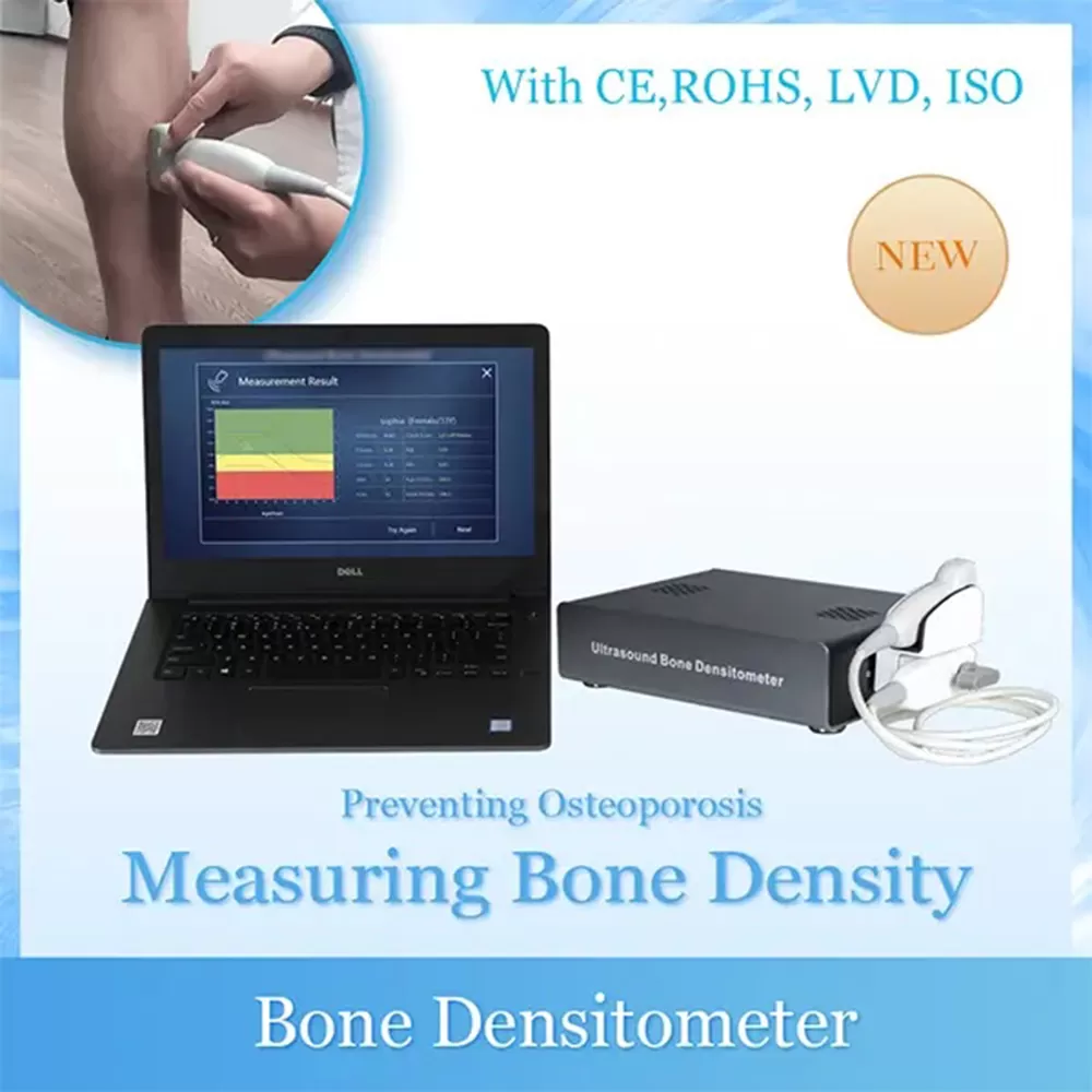
Portable Ultrasound Bone Densitometry BMD-A1 Osteoporosis Detector
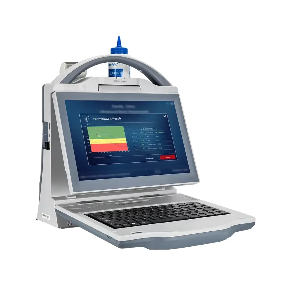
Portable Ultrasound Bone Densitometer BMD-A3
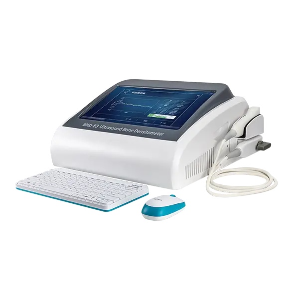
Automatic High Effective Portable Ultrasound Bone Densitometer BMD-B3
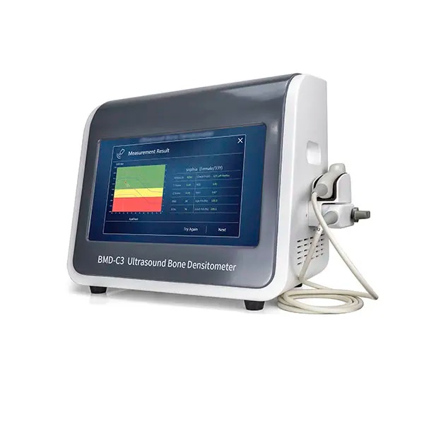
Portable Ultrasound Bone Densitometer BMD-C3
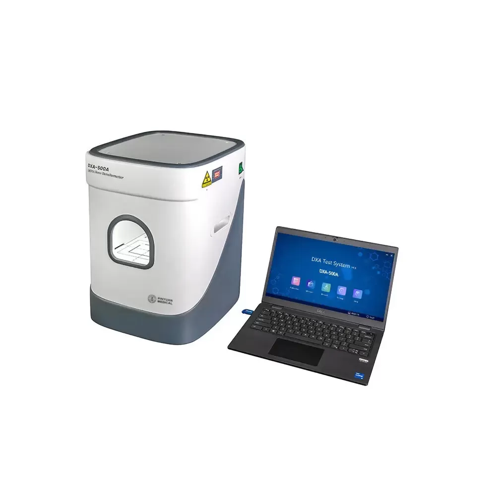
DXA Bone Densitometer 500A Dual-Energy X-ray Absorptiometry
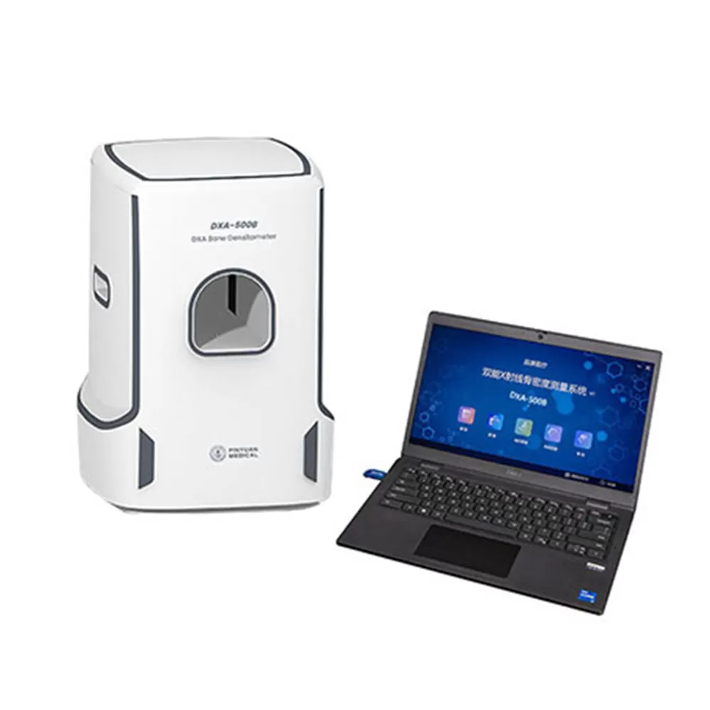
Dual-Energy X-ray Bone Densitometer DXA-500B
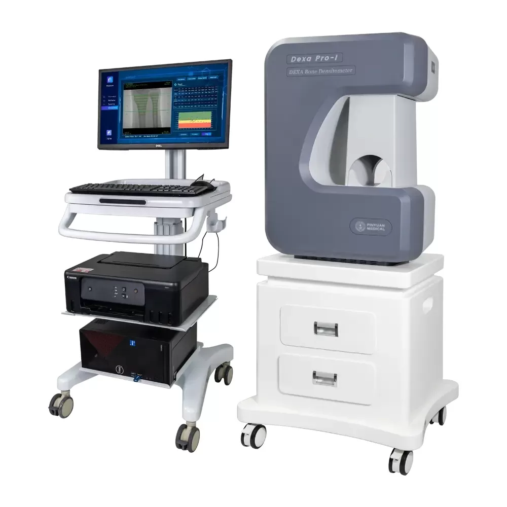
DXA Bone Densitometer DEXA Pro-1
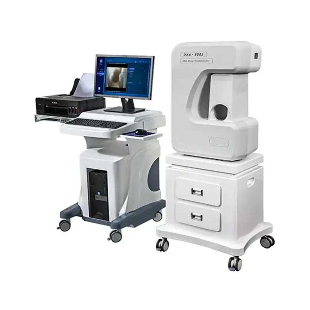
DXA Bone Densitometer 800E
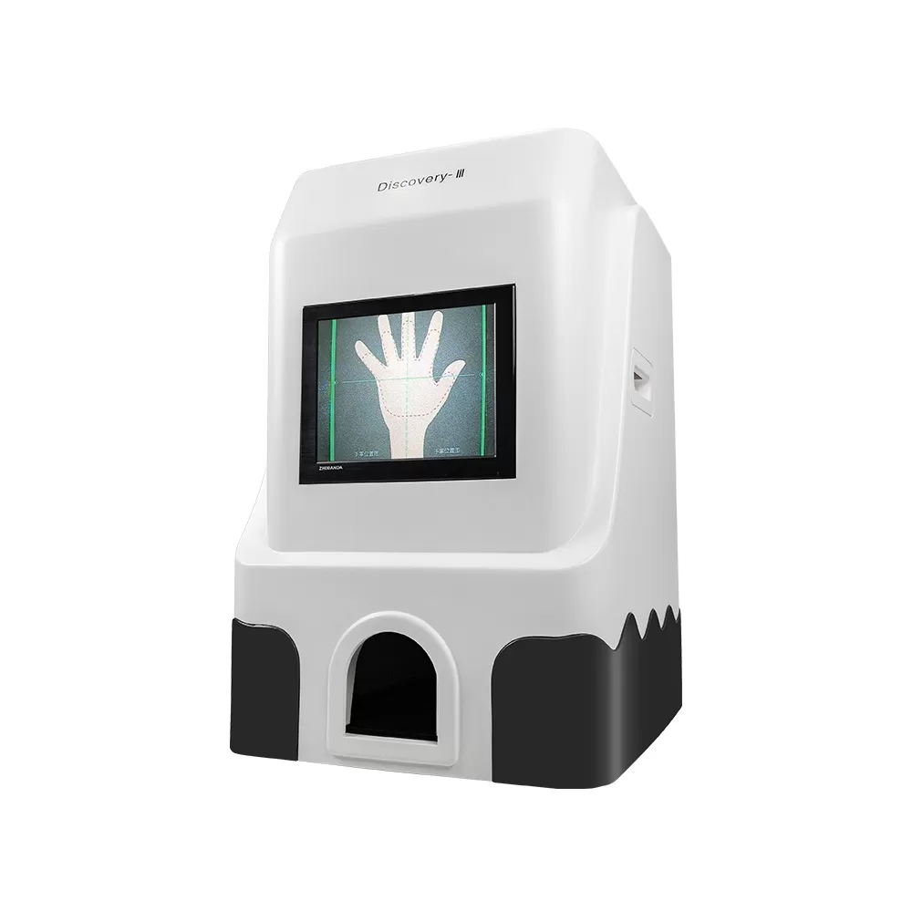
X-ray Bone Density and Skeletal Age Instrument Discover-lll